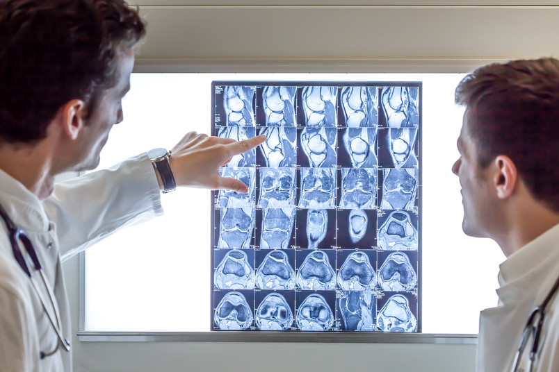
More precise diagnostics with medical imaging techniques
Statisticians at Ruhr-Universität Bochum have secured funding for a new project from the German Federal Ministry of Education and Research. They want to further develop the mathematical techniques needed to interpret data from medical imaging procedures. This should enable more precise diagnostics in the future. The team led by Dr. Nicolai Bissantz and Professor Dr. Holger Dette from the Chair of Stochastics will receive around 190,000 euros over three years from the "Mathematics for Innovation in Industry and Services" program.
The work for the project is embedded in the joint project "Dynamic medical imaging: modeling and analysis of medical data for improved diagnosis, monitoring and drug development (Med4d)". The University of Münster is coordinating the joint project, which also involves the University of Lübeck and industrial partners.
Finding the smallest pathological changes
To search for the smallest tumors or for pathological changes in the spine, doctors use imaging techniques such as computed tomography or positron emission tomography. These techniques produce images from different sectional planes through the body. However, they do not image the structures inside the body like a photograph. Initially, they only provide information about tissue density or metabolic activity, from which the actual image must then be reconstructed. This is based on a mathematical method: the Radon transformation, named after the mathematician Johann Radon, who was active at the beginning and middle of the 20th century.
Important here is the regularization parameter, which introduces the signal-to-noise ratio - i.e. the accuracy of the data - into the image reconstruction. If one underestimates the accuracy of the data, small details are no longer recognizable in the reconstructed image. Overestimating it creates artifacts in the reconstructed image that can simulate a pathological change.
Enabling efficient diagnostics
The regularization parameter must be selected as accurately as possible for each individual image of a patient. Only then are optimal image reconstruction and efficient diagnostics possible. The Bochum mathematicians are developing methods for the optimal selection of this parameter. In this way, they want to help improve data evaluation in order to achieve the best possible results with the currently existing medical technology equipment.
Click here for the original article in the RUB news.
